
The retina and vision
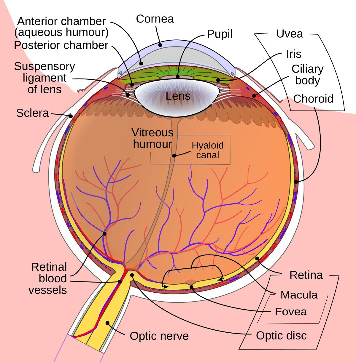
The retina is the thin, light-sensitive tissue that lines the inside of the back of the eye. It converts light into nerve signals that travel to the brain.
The macula is its central area. It allows us to read, recognize faces and see fine detail.
Arteries carry blood and oxygen to the retina. Veins carry blood away from it. Good circulation is essential for the retina to work.
What is RVO?
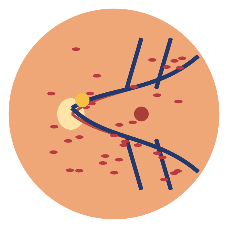
Retinal vein occlusion (RVO) happens when blood cannot drain properly, or at all, through a vein at the back of the eye. The blood pools, and bleeding (hemorrhages), fluid leakage and a lack of oxygen can occur.
If the fluid collects in the macula, it causes swelling (edema) and reduces central vision. The condition usually affects one eye and is not contagious.
Two main types
Branch retinal vein occlusion (BRVO)
Affects a branch of the vein and affects part of the retina. The effect on vision depends mainly on whether the macula is involved.
Central retinal vein occlusion (CRVO)
Affects the main vein and can affect a larger area. There is also hemiretinal vein occlusion, which affects about half of the retina.
Each type can come with a mild or a severe disturbance of the blood supply. Ischemia means that the tissue does not receive enough blood and oxygen.
Symptoms
Most often there is painless blurring or reduction of vision in one eye. The onset can be sudden, or vision can get worse over hours or days.
You may notice distorted lines, a dark spot, difficulty reading, or a part of the visual field that looks blurred. Some small branch occlusions are found by chance.
Any new, sudden loss of vision needs immediate medical assessment. We cannot tell the cause from the symptoms alone.
New, sudden drop in vision? Seek immediate medical assessment.
Risk factors
High blood pressure, raised cholesterol, diabetes, smoking and glaucoma are linked with an increased risk. The condition becomes more common with age, but it can also occur in younger people.
Your general health needs to be checked together with your personal doctor. In younger patients, when both eyes are affected, or when the picture is unusual, more targeted investigation may be needed.
Tests for thrombophilia (a tendency to form blood clots) are not needed for everyone. They are chosen based on your medical history and the findings.
How is it diagnosed?
Clinical examination
We measure your vision and eye pressure and examine the fundus (the back of the eye) with the pupil dilated. We check the macula, any hemorrhages and any new vessels.
OCT (optical coherence tomography)
OCT gives detailed cross-sections of the retina. It shows the edema and helps us assess the response to treatment.
Fundus photography
Records the extent of the changes and allows comparison at later visits.
Special examinations
Fluorescein angiography
Using photographs taken after a dye is injected into a vein, we examine the circulation, any leakage and areas with reduced blood supply. It is done when it will help with treatment decisions.
OCT angiography
Images the flow in the small vessels without injecting a dye. It complements the examination, but it does not show leakage and does not always replace fluorescein angiography.
General check-up
Usually includes blood pressure and tests for blood sugar, blood fats (lipids) or other factors, depending on your medical history.
Treatment of the edema
Intravitreal anti-VEGF injections
These are the main treatment option for macular edema that affects vision. The drug is given into the eye under local anesthesia and reduces leakage from the vessels.
Repeated injections are usually needed, at short intervals at first. After that, treatment is adjusted according to your vision and the OCT. There is no set number of injections that applies to everyone.
Corticosteroid implant
Used in selected cases. It can raise the pressure inside the eye and speed up the development of cataract, so appropriate monitoring is needed.
Laser and other interventions
When is laser needed?
If abnormal new vessels appear, laser treatment (photocoagulation) of the ischemic areas may be needed. The aim is to reduce the risk of bleeding and of neovascular glaucoma.
In selected branch occlusions, laser can also play a role in treating the edema. Not all patients need laser.
What does treatment not do?
The injections mainly reduce the edema; they do not mechanically remove the blockage. Aspirin or anticoagulants are not started just to treat RVO, without another medical reason.
Prognosis and follow-up
The course differs from person to person. It depends on the type of occlusion, the vision at the start, the blood supply to the macula and how long the edema has been present. Improvement can take months and is not always complete.
Check-ups continue even when vision improves, because the edema can come back or new vessels can appear. Ischemic CRVO needs especially close monitoring.
The interval between visits is set individually. Do not cancel injections or check-ups because the eye seems better.
In your daily life
Keep your blood pressure, blood sugar and cholesterol under control. Stop smoking and follow your personal doctor's advice on diet and exercise.
Reading and screens do not make the occlusion worse. Good lighting and low-vision aids can help. Whether you can drive depends on your vision and visual field.
Severe pain, redness or a sudden drop in vision need immediate examination. After an injection, increasing pain or reduced vision require urgent contact with an ophthalmologist.
Sources
- Royal College of Ophthalmologists. Retinal vein occlusion guideline (2022). doi.org/10.1038/s41433-022-02007-4
- Cambridge University Hospitals. Retinal Vein Occlusion, patient information (2025). cuh.nhs.uk/patient-information/retinal-vein-occlusion
This is patient information. Examinations and treatment are tailored to each patient by the treating doctor.