
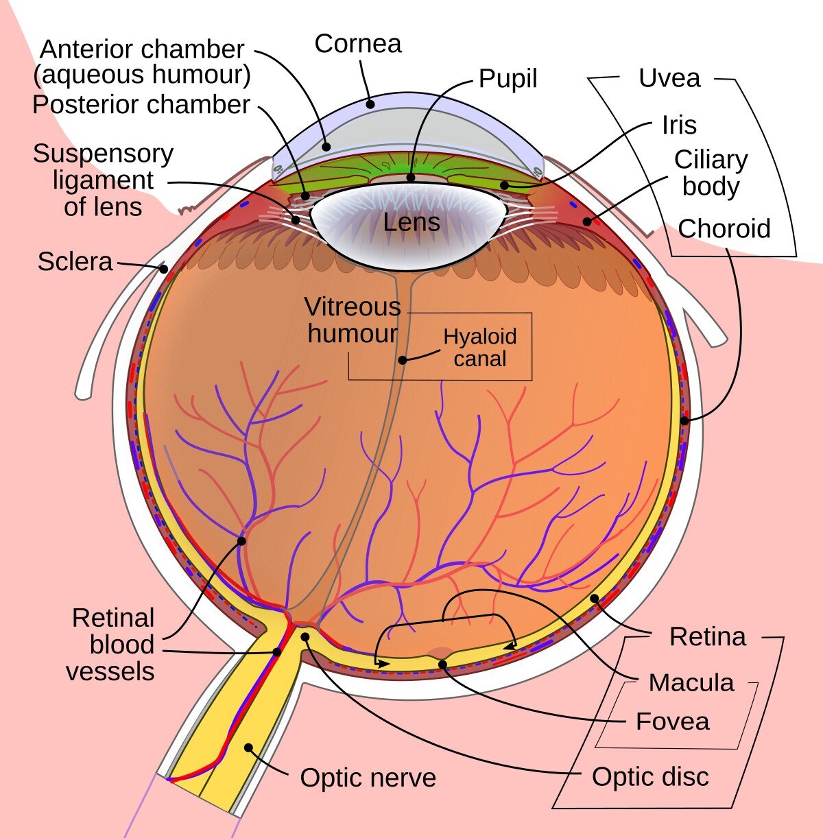
The retina and vision
The retina is the thin, light-sensitive tissue that lines the inside of the back of the eye. It converts light into nerve signals that travel to the brain.
The macula is its central area. It allows us to read, recognize faces and see fine detail.
Arteries carry blood and oxygen to the retina. Veins carry blood away from it. Good circulation is essential for the retina to work.
What is RAO?
Retinal artery occlusion (RAO) happens when the supply of blood and oxygen to part or all of the retina is blocked. It can cause permanent damage to vision.
It is often caused by an embolus, that is, material carried in the blood from another vessel or from the heart. In some cases there is inflammation of the artery walls.
Acute retinal artery occlusion is treated as a vascular emergency. It needs immediate investigation, also to protect your general health.
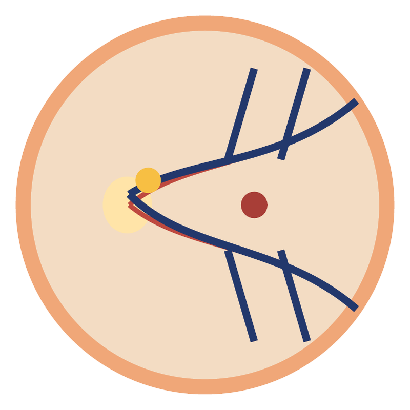
Two main types
Central retinal artery occlusion (CRAO)
The main artery of the retina is blocked. It usually causes sudden and significant loss of vision in one eye.
Branch retinal artery occlusion (BRAO)
A smaller branch is blocked. Part of the visual field may be lost, while central vision may be preserved if the macula is not affected.
Both forms need urgent vascular assessment. Seeing well in the center does not rule out a significant arterial occlusion.
Symptoms and immediate action
The typical symptom is a sudden, usually painless loss of vision, or a dark area appearing, in one eye. It can happen within seconds.
Even if vision comes back after a few minutes, the temporary loss may be a warning vascular event and needs immediate assessment.
Call 112 or go straight to an Emergency Department. Note when the symptoms started, or when you last saw normally. Do not drive and do not wait for your next appointment.
Sudden loss of vision: call 112. Every minute counts.
Why do we check the whole body?
The occlusion may be linked to atherosclerosis of the carotid arteries or to a source of emboli in the heart, such as atrial fibrillation. It may be a sign of an increased risk of stroke.
Risk factors are high blood pressure, diabetes, raised cholesterol, smoking and cardiovascular disease.
New weakness or numbness on one side of the body, an uneven (drooping) face or difficulty speaking require an immediate call to 112. Do not wait for them to pass.
Treatment in the acute phase
The priority is immediate transfer to a hospital that can assess strokes. The eye examination and the vascular work-up are organized without unnecessary delay.
In some patients with a very recent CRAO, the specialist team may consider reperfusion therapy (treatment to restore blood flow), depending on when the symptoms started, any contraindications and the protocol in use. It is not suitable for everyone and does not guarantee that vision will recover.
There is no treatment that reliably restores vision in every case. Delay reduces the options available.
When there is arteritis
Giant cell arteritis
In people usually over 50, an inflammation of the arteries can also threaten the vision of the other eye.
Report immediately any new headache, tenderness of the scalp, jaw pain when chewing, fever, weight loss, or pain and stiffness in the shoulders and hips.
Urgent blood tests and further investigations are carried out. If the clinical suspicion is strong, the doctor starts corticosteroids immediately, without waiting for final confirmation. Do not take any treatment on your own.
Eye examinations
Vision and fundus examination
We check visual acuity, the pupils, the eye pressure and the retina. There may be whitening of the area that is not receiving blood, or a visible embolus inside an artery.
OCT (optical coherence tomography)
In the acute phase, OCT can show swelling and increased reflectivity of the inner retinal layers. Later, thinning of the tissue may be seen.
Visual field and angiography
These are used as needed to record the damage and the circulation. They must not delay urgent referral.
Vascular and cardiac work-up
The hospital team decides which tests are needed. They may include imaging of the brain and of the blood vessels of the head and neck, as well as a check of the carotid arteries.
An electrocardiogram (ECG) and, when indicated, longer heart-rhythm monitoring or an ultrasound of the heart help in the search for a source of emboli in the heart.
Blood pressure, blood sugar, blood fats (lipids) and other blood values are checked. Special tests are chosen based on age, medical history and the likely cause.
Prognosis and follow-up
The prognosis depends on the vessel involved and on the extent and duration of the ischemia. In CRAO, the loss of vision is often severe and permanent. In BRAO, central vision may be good, but a gap in the visual field may remain.
Follow-up visits record vision, eye pressure and any complications, such as abnormal new vessels. The schedule is adjusted to the findings and is not the same for everyone.
Monitoring of the eye continues alongside treatment of the vascular cause.
Preventing another episode
Treatment depends on the cause. It may include an antiplatelet drug, a statin or, in certain heart conditions, an anticoagulant. The choice is made by the responsible medical team.
Keep your blood pressure, cholesterol and blood sugar under control, stop smoking, and follow the advice on exercise and diet. Do not start or stop aspirin or an anticoagulant on your own.
Low-vision aids and adjustments to daily life can help. Fitness to drive needs an assessment of your vision and visual field.
Sources
- American Heart Association. Management of Central Retinal Artery Occlusion (2021). doi.org/10.1161/STR.0000000000000366
- American Academy of Ophthalmology. Retinal and Ophthalmic Artery Occlusions, Preferred Practice Pattern. aaojournal.org
This is patient information. Examinations and treatment are tailored to each patient by the treating doctor.