Vision and the retina
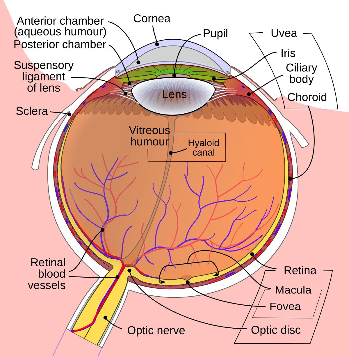
Vision is one of our most important senses. When we open our eyes, light enters the eye. It passes through the cornea, the lens and the vitreous and is then absorbed by the retina.
The retina consists of millions of tiny visual cells (photoreceptors) and is responsible for converting the visual image into an electrical signal, which the brain then processes and turns back into an image. In other words, it works as a kind of film that records what we see.
In the center of the retina there is an area of 2–3 mm called the macula. It is an extremely sensitive area, responsible for sharp vision (the details of objects) as well as for color perception. Disease-related changes that occur here are therefore especially serious.
Diabetes and the eye
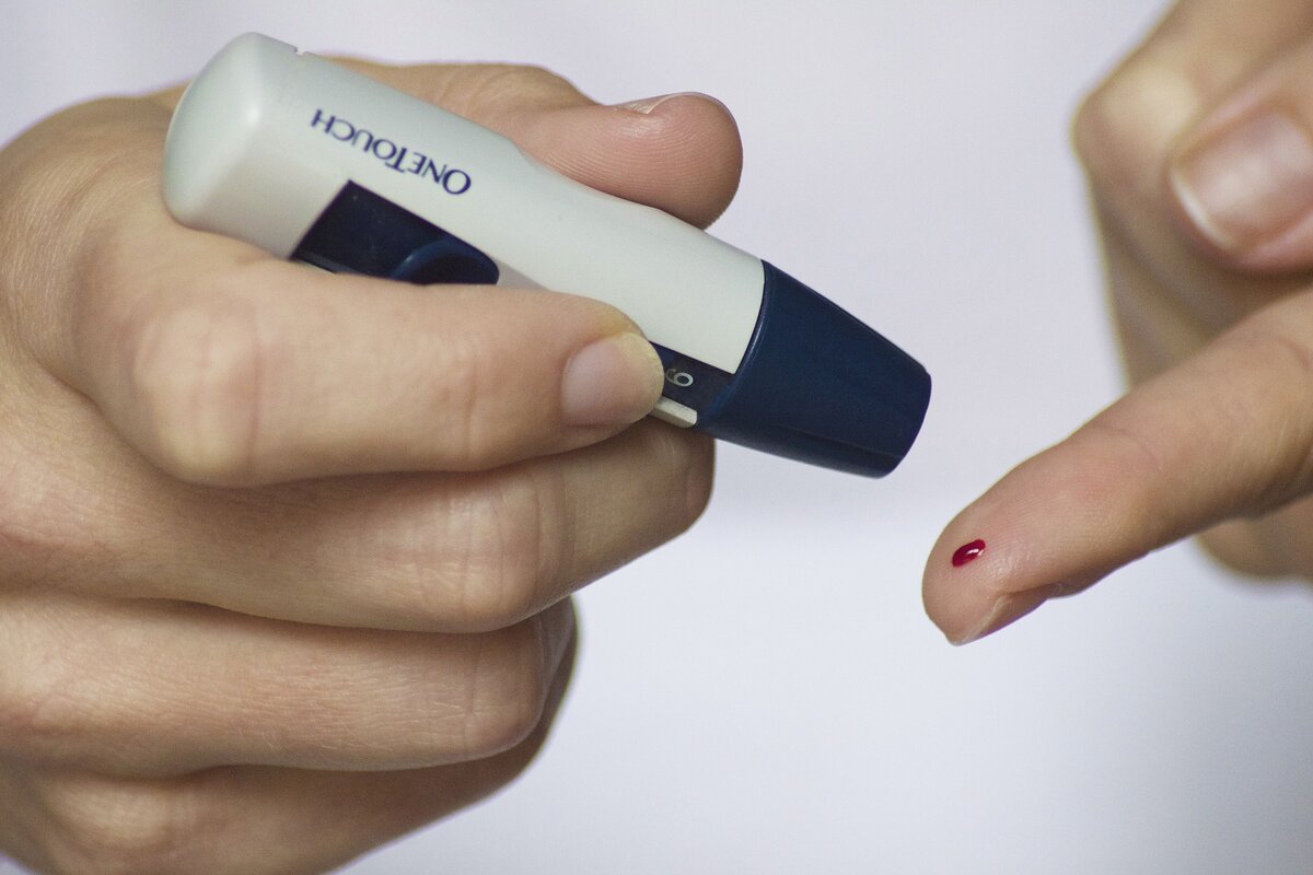
Diabetes affects your body in many different ways. Among other things, changes can occur in the body’s blood vessels. When diabetes affects the blood vessels in the retina of the eye, this is called diabetic retinopathy.
Diabetic retinopathy is insidious: the changes are completely symptom-free at first, meaning that visual acuity (vision) remains normal. Gradually, however, the central part of the retina, the macula, may become affected. Your visual acuity is then affected. Bleeding inside the eye may also occur, leading to a sudden worsening of vision.
By examining your fundus regularly, we can detect changes early, before symptoms begin to appear. This is the most important point, because treatment usually only slows the course of the disease. Visual acuity that has been lost in one eye cannot always be regained.
What happens in the eye?
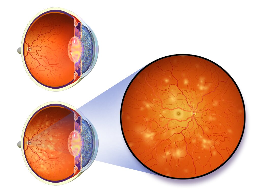
The retina consists of millions of visual cells and a large number of tiny blood vessels. In diabetes, raised blood sugar levels lead to metabolic disturbances that weaken the vessel walls and cause small bulges to form in these tiny blood vessels.
These bulges are the first visible changes, and on examination they appear as small red dots on the retina (microaneurysms).
In some cases, the bulges can burst and leak through their walls. Changes then appear in the form of swelling (edema), bleeding, or leakage of blood fats (exudates). When the swelling or the exudates reach and affect the macula, the retina’s ability to reproduce visual impressions is disturbed.
At a later stage, the tiny blood vessels can become blocked, which leads to poorer blood flow and a lack of oxygen in the retina. The retina tries to compensate for the lack of oxygen by forming new blood vessels. These are weak and break easily, causing bleeding that can lead to a rapid worsening of vision.
Eventually, the abnormal new blood vessels can pull on the retina (traction), which then risks becoming detached. In the worst case, this can cause blindness.
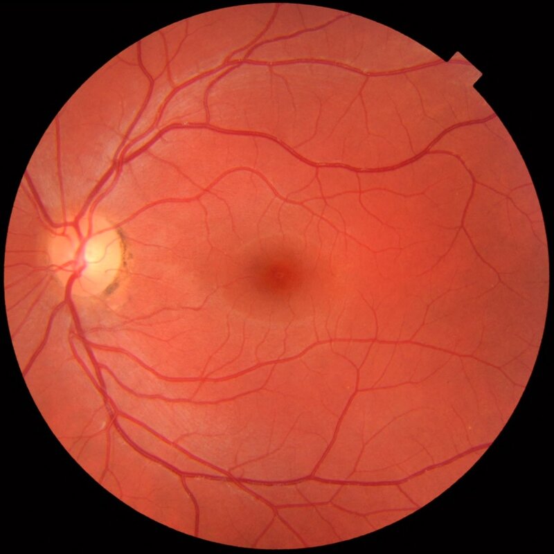
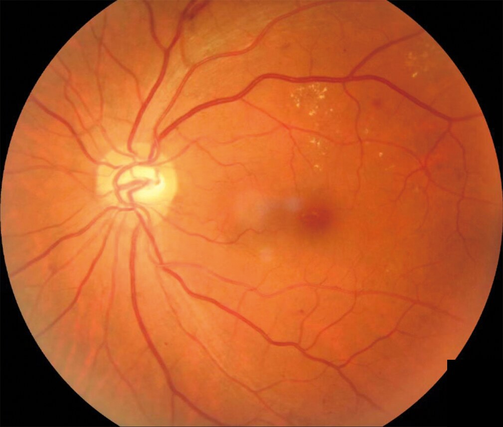
Diabetic retinopathy and its stages
About one third of all people with diabetes have some form of retinal changes. Of these, 10–35% are at risk of losing vision.
In people with type 1 diabetes who developed the disease before the age of ten, there are practically no retinal changes before puberty. In people with type 2 diabetes, retinal changes are already visible in up to one third of cases at the time the patient is diagnosed with diabetes.
Two forms of sight-threatening retinal changes
In people with type 1 diabetes, the growth of new small blood vessels is the most common form. In people with type 2 diabetes, changes in the area around the macula are more common.
Stages
There is a non-proliferative stage and a proliferative stage.
- Non-proliferative stage: divided into mild, moderate and severe. At these stages you do not need treatment.
- Proliferative stage: abnormal new vessels are present. These vessels grow because of a factor called VEGF, which is produced when an area of the retina becomes ischemic, that is, does not receive enough blood. Because these vessels are abnormal, they bleed and leak, and if no treatment is given there is a risk that they lead to retinal detachment.
Risk factors
There are certain factors that we know affect how quickly and how severely diabetic retinopathy develops. The most important risk factors are:
- Duration: how long someone has had diabetes. After 20 years with the disease, about 90 percent of both type 1 and type 2 patients have some form of retinal changes.
- Poor control of blood sugar levels. The blood sugar level is of decisive importance in both type 1 and type 2.
- High blood pressure. Blood pressure should not exceed 130/80 mmHg.
- High levels of blood fats. Certain raised blood fat values can contribute to retinal changes.
- Hormonal changes in the body. Pregnancy, for example, can accelerate retinal changes.
Prevention & screening
If you have diabetes, you can help prevent changes in the retina. This requires good control of your diabetes, with low and stable blood sugar levels, good blood pressure and good blood fat levels.
Go for regular check-ups with your ophthalmologist to have photographs taken of the back of your eyes (diabetes screening). In this way, we can detect small changes that are completely symptom-free, early and at an initial stage.
Fundus photography (diabetes screening)
We photograph your fundus to keep a record and to be able to monitor any changes over time. To photograph your fundus with the best possible image quality, we give you eye drops that dilate the pupil and make it larger. After about half an hour, or when the pupils are large enough, we can photograph your fundus with a special camera.
In most European countries, systematic fundus photography for people with diabetes is required by the health system and is called screening.
First examination
If you have type 1 diabetes, you should go for regular check-ups from the age of ten. If you have type 2 diabetes, you should have your eyes examined when you are diagnosed with diabetes.
Regular check-ups
Type 1 diabetes
- No retinal changes and no risk factors: a new check-up every 2 years.
- Diabetic retinopathy: a check-up every 3 months to 1 year, depending on its degree.
Type 2 diabetes
- No retinal changes and no risk factors: a new check-up every 3 years.
- Diabetic retinopathy: a check-up every 3 months to 1 year, depending on its degree.
Pregnancy (type 1 or 2)
- A check-up during the first trimester of pregnancy.
Special examinations
If the examination shows that there are changes in the fundus, or that there are certain risk factors that can affect the speed and severity of the changes (for type 1 and type 2), we adjust the intervals between check-ups to suit you. There are a few different types of examination.
Vision test
We test your vision using a standard letter chart.
OCT examination (optical coherence tomography)
In case of swelling and leaking blood vessels in and near the macula, we can examine the retina with an OCT examination. We do this so that we can study the different layers of the retina. To make the examination easier, you are given eye drops that dilate the pupil. After about half an hour, or when the pupils are large enough, we can take a cross-sectional image of the retina. With the help of the OCT images, we can see whether your eye has abnormal changes in the retina and the macula.
Fundus fluorescein angiography
This is a painless examination performed by injecting a special dye into a vein. When the dye reaches the eye and the retina, we photograph the fundus with a fundus camera. We carry out this examination in case of more serious changes in the eye, so that we can examine the vessels and the circulation in the fundus before possible laser treatment.
Ocular ultrasound
In case of severe bleeding that makes it difficult to see the back of the eye, we examine the retina using ultrasound.
Treatment
Laser photocoagulation
Traditionally, severe retinal changes are treated with laser during a visit to the doctor. The type of laser treatment we perform depends on the type of changes you have. If the problem is newly formed blood vessels growing on the retina, we perform so-called panretinal photocoagulation (PRP), which is an extensive laser treatment of the retina. The treatment slows the formation of new blood vessels and reduces the risk of serious complications such as bleeding and retinal detachment.
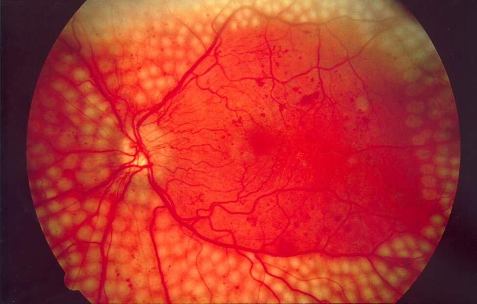
Intravitreal injections
If the changes are close to the macula, they can often be treated with a mild laser. If, on the other hand, the change is in the center of the macula, we cannot use a laser, as it risks damaging the macula. Instead, we treat the changes with injections into the eye. The injections are given into the eye under local anesthesia. With the injections we halt the progression of the condition and of the macular edema, and vision often improves. The treatments usually need to be repeated and require regular check-ups.
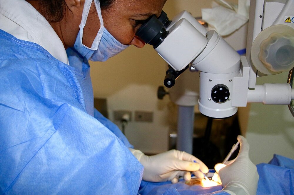
Vitrectomy
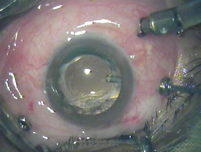
In case of more severe changes in the retina, we can perform surgery on the vitreous body, so-called vitrectomy. During the operation we remove the vitreous body and any blood present. Then, if necessary, we can operate on the retina. We perform this type of operation if:
- There is a large amount of bleeding in the vitreous, clouding your vision.
- There is traction on the retina, which then risks becoming detached.
Depending on the type of changes and whether treatment is started early, we achieve good results in 50–90% of cases.