
The retina and how the eye works
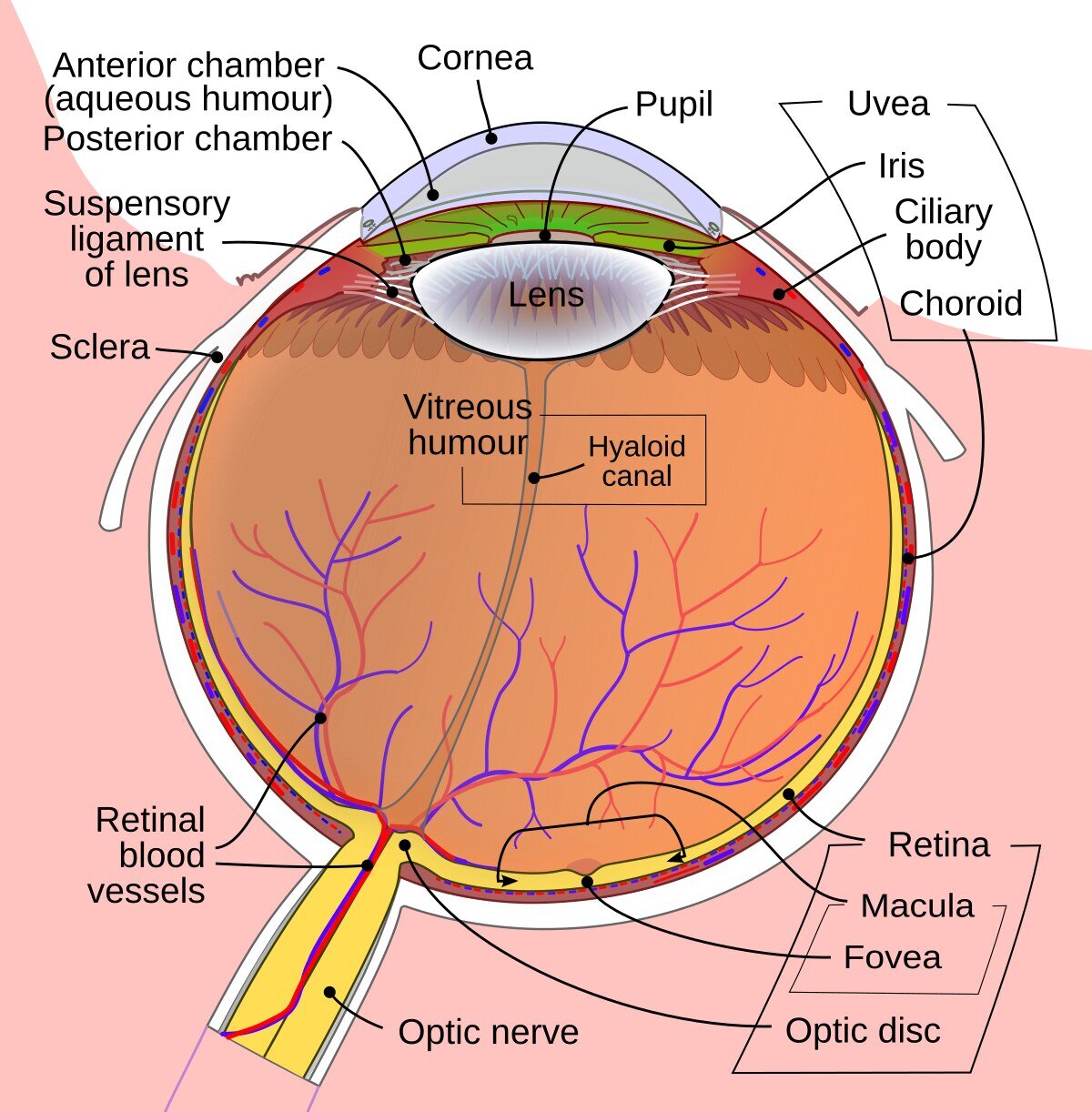
The retina is a thin membrane that lines the inside of the eyeball and contains the light-sensitive cells that convert incoming light into nerve signals. You can compare the retina to the film in a camera.
The macula is the area in the middle of the retina where most of the visual cells are located and where you see most sharply and clearly.
The choroid is a membrane rich in blood that lies between the white of the eye and the retina. It supplies the retina with oxygen and nutrients and removes waste products that are no longer needed.
What is CSCR?
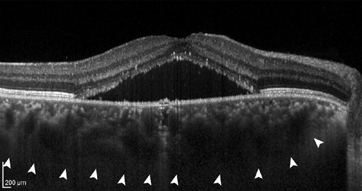
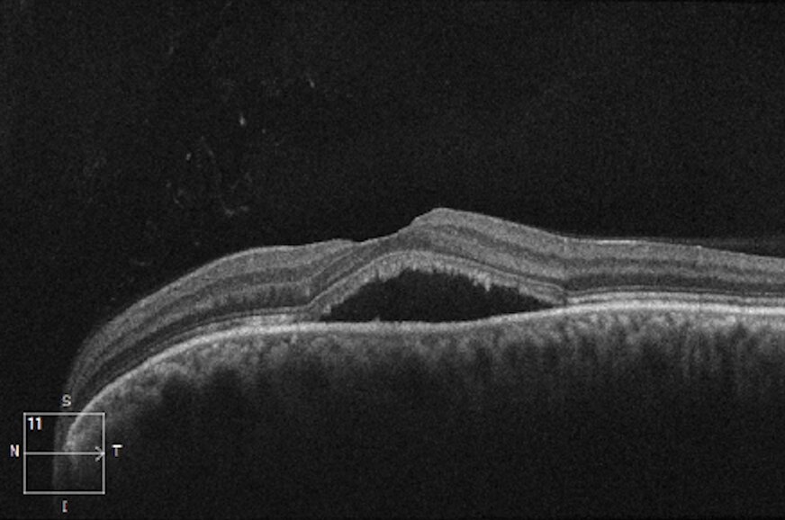
Central serous chorioretinopathy (CSCR) is a disease in which fluid has collected under the retina, causing swelling (edema) of the macula. The disease usually appears as a visual disturbance: a round spot or blurred area in the center of the visual field, while colors and the size of objects are perceived as distorted (metamorphopsia).
Most patients have a thick choroid, because the normal outflow of blood from the choroid into the circulation is obstructed. This causes the choroidal vessels to dilate; the pressure inside the vessels rises and the vessel walls let through more fluid than normal. If the barrier between the choroid and the retina is also damaged at one or more points, fluid can leak underneath the retina.
Most people who develop the disease are of working age (20–50). Men are affected more often than women (five times as often). Women are usually about ten years older than men when they are affected. In about 85% of cases the disease resolves on its own, but those with the more severe variant of the disease may be left with permanent visual impairment. However, the disease does not lead to blindness.
Acute and chronic CSCR
There are several ways of classifying the disease. The most common is to distinguish between acute and chronic CSCR.
Acute CSCR
The patient usually seeks help because a spot or some other form of visual disturbance has suddenly appeared in the center of the visual field. It is common for the patient to see colors differently and to perceive objects as smaller compared with the healthy eye. The disease heals on its own within 3–6 months in about 85 percent of cases. Up to half of all patients are affected more than once.
Chronic CSCR
When the fluid has not been absorbed within 3–6 months, the disease is called chronic and must be treated. There is also a group of patients who visit their ophthalmologist for the first time because symptoms have developed gradually, with no earlier symptoms, but who already show signs of chronic CSCR with more extensive retinal damage at the first visit. In this type of more severe chronic CSCR, we may decide on treatment already at the first visit.
Although symptoms are more often noticed in one eye, up to 40% of all patients have signs of the disease in both eyes. Patients over 50 are more likely to have the disease in both eyes (over 50% of cases).
Risk factors
Cortisone
The most important external risk factor for CSCR is the use of cortisone (corticosteroids). Even small amounts of cortisone, for example from nasal sprays, inhalers or skin creams, appear to matter. We recommend that patients stop or postpone using cortisone if possible.
Stress
Another risk factor is stress. The condition appears to be directly linked to stress, particularly in patients with a “Type A” personality (ambitious, workaholic). These people are often in a hurry and competitive. They take on a lot and often do many things at the same time. Studies show that these patients, on average, have higher levels of cortisol (the stress hormone) in their blood, which may explain the link with the disease.
Other factors
Shift work, poor sleep, pauses in breathing at night (sleep apnea) and pregnancy also mean an increased risk of developing the disease.
Examinations
There are many different examinations we can carry out to make a diagnosis.
Visual acuity test
We measure visual acuity both near and far. The measurement shows how much your vision and your ability to make out small details have been affected.
Eye examination under a microscope
We look at the retina and examine the macula with a special microscope. Sometimes it is possible to see whether there is swelling in the macula, whether there are extensive changes indicating long-standing disease, or signs of another eye disease that could explain the fluid accumulation (for example, leaking vessels due to age-related macular degeneration).
OCT (optical coherence tomography)
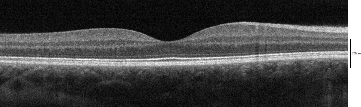
During an OCT examination, we take cross-sectional images of the retina so that we can see details at very high magnification. With the help of the images, we can study the optical properties of the fluid to find out how long the disease has been present.
OCT angiography
Sometimes we use OCT angiography to image the retinal vessels and rule out that the swelling under the macula is caused by leakage from newly formed abnormal vessels due to age-related macular degeneration, because that condition needs immediate treatment with intravitreal injections. This examination does not require an injection of contrast dye.
Fundus angiography
This is a painless examination performed by injecting a special dye into a vein. When the dye reaches the eye and the retina, we photograph the back of the eye (the fundus) with a fundus camera. In this way, we can locate areas that need treatment.
Autofluorescence
By photographing the fundus with a special light, we can see both overactive and inactive parts of the retina. This helps us assess how widespread the disease is and whether it is chronic or acute.
Treatment
The aim of treatment is to remove the fluid in order to protect the visual cells and prevent permanent damage. Many types of treatment have been proposed over the years and are still used in many hospitals around the world. However, there is a lack of good research confirming the effectiveness of these treatments. The only treatment supported by high-quality scientific research is photodynamic therapy (PDT).
Photodynamic therapy (PDT)
PDT is performed by injecting a special light-sensitive substance into a vein. We then illuminate the active areas of the retina and choroid with light of a specific wavelength, which activates the light-sensitive substance and acts on the diseased blood vessels.
In about 80% of patients, the swelling decreases with PDT. The treatment may need to be repeated. For patients with long-standing chronic CSCR, there is a risk that the treatment will not be sufficiently effective.
In acute CSCR, treatment usually leads to full recovery of visual acuity, although the patient may notice minor visual disturbances such as reduced contrast and altered colors. Treatment of chronic CSCR reduces the risk of recurrence and stabilizes or slows the progression of the disease. Usually, visual acuity does not return to what it was before.
Treatment of acute CSCR
Since acute CSCR usually heals on its own, we do not need to treat it immediately; instead, a follow-up at 3 months is recommended. If there is still fluid under the retina three months after the disease appeared, and we judge it to be a threat to vision, we treat the disease with photodynamic therapy.
Treatment of chronic CSCR
Because patients with chronic CSCR have a worse prognosis, we can carry out photodynamic therapy already at the first visit. However, the amount of fluid under the retina varies and sometimes disappears completely, which means that we sometimes choose to wait.
Anti-VEGF injection
In some cases of chronic CSCR, the swelling of the macula is caused not only by a malfunctioning choroid but also by newly formed, fragile blood vessels under the macula that leak. We can then slow the formation of new vessels and prevent the leakage by injecting a drug (anti-VEGF) into the eye.
Other treatments
If the leakage points are far enough from the macula, we can seal the leaking vessels with a laser. This treatment is quicker than PDT, but it does not target the underlying cause of the disease (a choroid that does not work properly). In addition, there may be some risk that the treatment causes a small blurred spot in the visual field that can grow larger over time, which is why laser treatment close to the macula is avoided.
Many other treatments have been described over the years with a positive effect in a few patients. Since the disease heals on its own in a large proportion of patients, large, well-conducted studies with a control group are needed to prove that a proposed treatment has a real effect.
Research
Many expert groups have worked on clinical and genetic research into CSCR. This has led to greater knowledge about the treatment and the genetic background of the disease. Many studies are under way to clarify the cause of the fluid accumulation under the retina and to develop new treatments, especially for patients with the severe chronic type of the disease, which is harder to treat. I had the pleasure and the good fortune to take part in some of this research during my time in Sweden.