

The macula
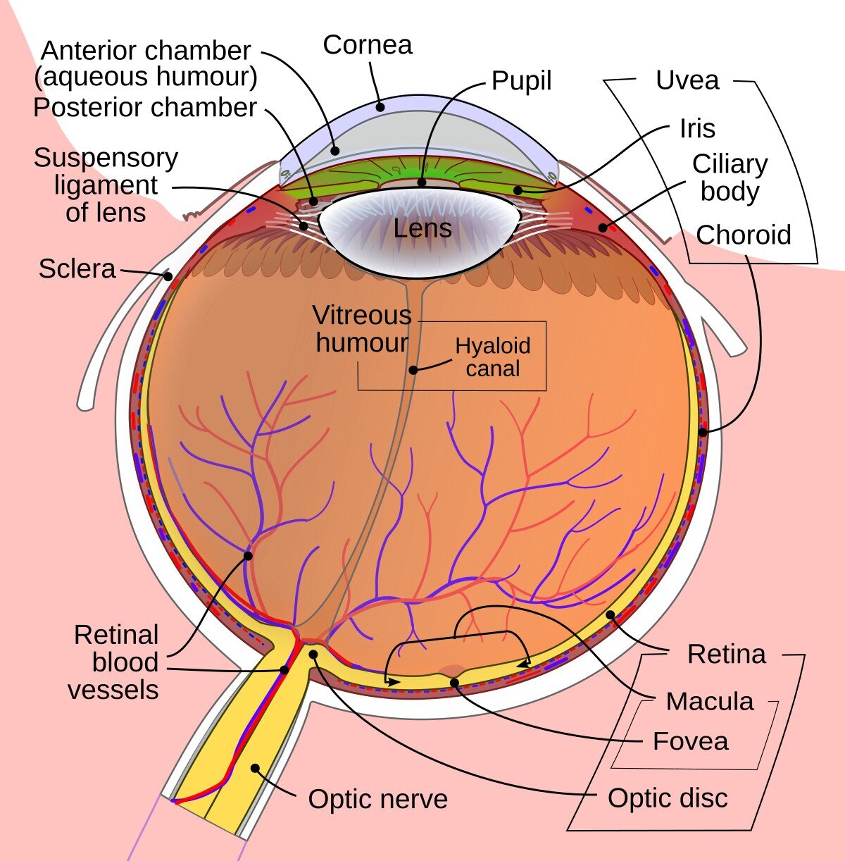
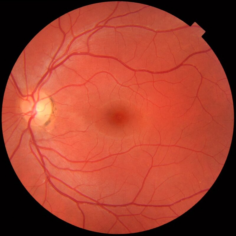
The macula is the area in the center of the retina where most of the visual cells are located, and it is responsible for sharp vision (the details of objects) as well as for color perception. This area is 2–3 mm in size and particularly sensitive to various kinds of damage. Any damage to the macula results in a loss of central vision, which is usually noticed immediately.
What is age-related macular degeneration?
Age-related changes in the macula are called macular degeneration. The changes affect visual acuity (vision). If you are affected, you will therefore have problems with sharp vision, that is, with reading and seeing small details. However, the changes do not greatly affect the visual field, your peripheral vision. You will therefore not go blind from age-related changes in the macula. Most people who develop the disease can continue to live an active and normal life.
The older you get, the more likely you are to have changes in the macula. The disease is the most common reason why people over 60 have reduced vision. The average age of those who receive treatment is 80. Women are at slightly higher risk of being affected than men, but this is mainly because women live longer than men. The disease is also more common in certain families. Smoking is a factor that affects the risk of developing the disease.
Dry and wet forms
There are two different forms of age-related macular degeneration: the dry form and the wet form.
Dry form
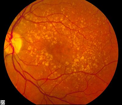
The dry form (also known as the non-exudative or atrophic form) is the most common and occurs in about 80% of cases; nevertheless, it is less dangerous. With dry changes, there is aging, thinning and atrophy of the macula, and vision gradually gets worse. Symptoms first appear in one eye and later in the other eye as well. Dry changes eventually always affect both eyes and are chronic.
Wet form
The wet form (also known as the exudative form) is much rarer than the dry form, but the disease can develop quickly. In the wet form, new vessels form behind the retina (neovascularization). These vessels are abnormal and very fragile, which causes blood and fluid to leak under the macula, which then swells. The swelling destroys the light-sensitive cells of the macula and your vision gets worse. The deterioration can happen within just a few weeks or months. It is therefore important to start any treatment as soon as possible.
Symptoms
The symptoms of age-related macular degeneration can vary slightly. You may see small spots in the center of your visual field, or you may find that everything looks gray and blurry. Many people find it harder to recognize faces. Letters often seem to move when reading a longer text. If you have changes in vision in only one eye, you may sometimes find it difficult to judge depth and distances.
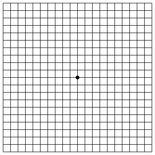
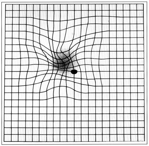
You may also notice that images are distorted: lines that are actually straight become curved, and faces can look distorted (known as metamorphopsia). This is more common in those who have the wet form. In long-standing disease, central vision can be lost completely, and you may see only a blurred dark spot in the middle of your visual field.
You can check for metamorphopsia yourself at home using special Amsler grids, as shown below:
Treatment
Dry form
Currently, there is no effective treatment for the dry form.
In some cases, when there are deposits in the macula (drusen), your doctor may advise you to take extra vitamins, as this may reduce the risk of developing wet-form changes. There is no scientific evidence that vitamins have positive effects on the macula if you do not have drusen.
Wet form
The wet form is usually treated with injections into the eye (intravitreal injection).
You will receive a drug that slows the formation of new blood vessels under the macula and reduces the swelling. We give the drug into the eye with a fine needle. The procedure is short and hardly felt at all, because you receive anesthesia. Before the injection, we wash your eye with an antiseptic to reduce the risk of infection.
Injection treatment does not cure the disease but slows its progression. The injections therefore need to be repeated many times, and you will have regular check-ups with your ophthalmologist.
Living with reduced vision
Using your eyes as usual does not affect your vision, so you should not be afraid of, for example, watching television or reading.
- Move closer to the television if you have difficulty seeing the subtitles or on-screen text.
- Don’t hesitate to get separate reading glasses, because multifocal (varifocal) glasses have a very small reading area. Simple reading glasses from the pharmacy often work quite well. These cannot harm your vision.
- It is important to have good, directed reading light, and a magnifying glass is often a good aid.
- You can also take a short break and rest your eyes if they get tired.
If your vision becomes very poor and reading becomes very difficult, you may need visual aids.
Over time, your vision may deteriorate to such an extent that you no longer meet the legal requirements for driving. It is your ophthalmologist’s duty to inform you about this.
Examinations
To make a diagnosis, we need several examinations. We check your visual acuity (vision), new vessels in the retina and macula, and the circulation in the retina and macula.
To make the most accurate diagnosis possible, you will receive pupil-dilating drops before the examination. The doctor can then see the retina and macula more clearly. The drops make your vision slightly blurry for a few hours. You should not drive a car immediately after the examination. You may also be dazzled by bright lights, so it may be a good idea to wear sunglasses.
Visual acuity test
We measure visual acuity at both near and far distances. The measurement shows how much your reading ability and your ability to make out small details have been affected by the changes of age-related macular degeneration.
Eye examination under a microscope
The doctor looks at the retina and examines the macula with a special microscope. In this way, the doctor can see whether there are early stages of age-related changes, or whether there is blood in the macula, which suggests that you are affected by the wet form.
OCT examination (optical coherence tomography)
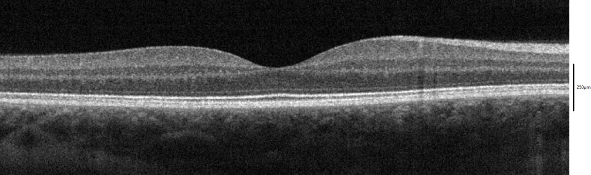
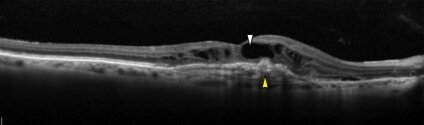
This is a non-invasive and completely painless examination technique. During an OCT examination, we take cross-sectional images of the layers of the retina. With the help of the images, we can see whether there is swelling in the macula. This may be a sign of neovascularization (wet form). We may then need to carry out another examination, OCT angiography, or a traditional angiography with contrast dye, to confirm the presence of the wet form. With angiography, we can see how blood flows in the retina.
Research
Research into age-related changes in the macula has been very intensive for several years. Above all, researchers are trying to find injectable drugs with a longer-lasting effect, so that the eye does not need treatment as often. Trials are also under way with injections that we hope can slow the dry form. I took part in several such studies during my time in Sweden, and we keep continuously up to date with new treatments.